
This was sent by Asad Tasleem, an Emergency Medicine resident in Pakistan:
"I am an ED Resident in a tertiary care Hospital Pakistan. I received a 70 year old male with DM, HTN who presented with history of fall of unknown mechanism possible syncopal episode. On arrival he was vitally stable and all of the attention was to the obvious deformity of hhis left leg which turned out to be a femoral neck fracture.
I ordered an EKG:

I sent it to the PMCardio Queen of Hearts AI ECG model. The Queen always asks about the pretest probability, because that is crucial to any test, and it is quite possible that an ECG which appears to represent OMI does NOT represent OMI if there is a very low pretest probability.

I answered "No" to the question.
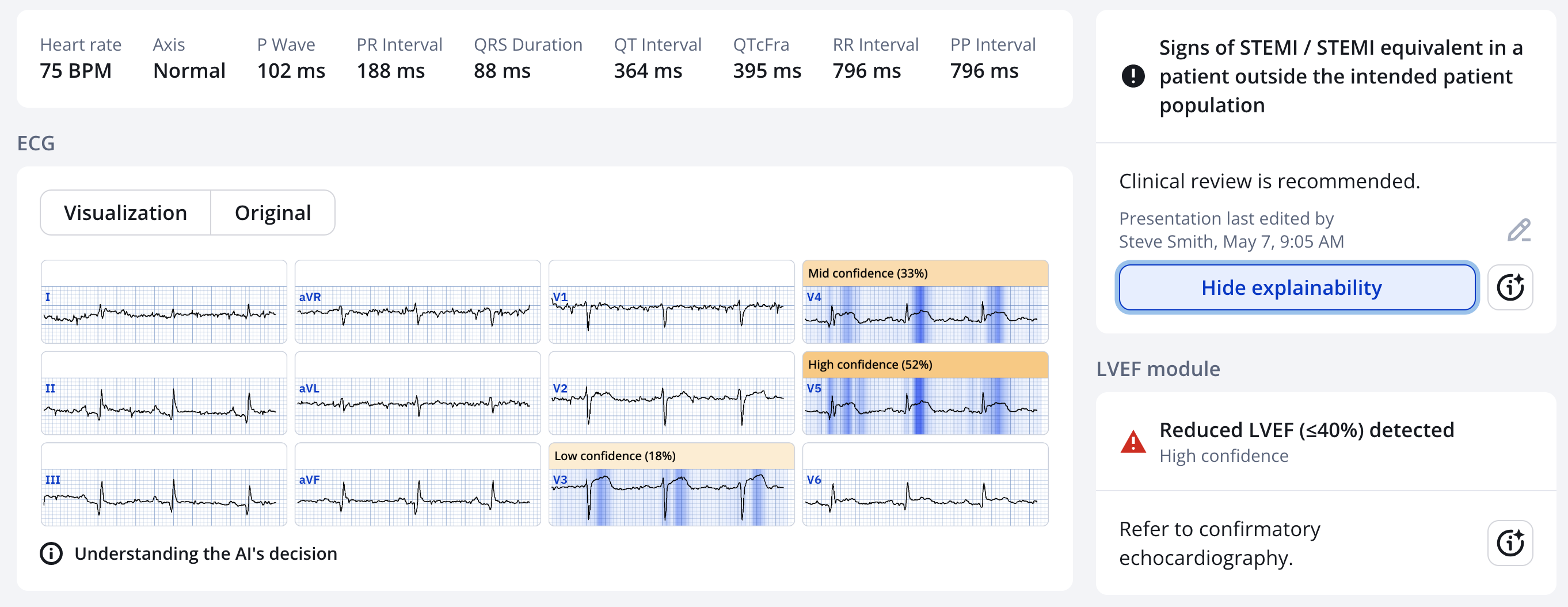
You can see that, in spite of the "No" answer, she is still worried about OMI
Asad continues:
"I thought it was very suspicious for Terminal QRS distortion in anterior leads. I rushed cardiology and as always they weren't convinced as the patient had no chest pain. Trop came positive and continue to rise on serial tests."
_____
Smith: strictly speaking, "terminal QRS distortion" is only proven to be highly specific for LAD OMI if it is in leads V2 or V3. However, it likely a significant finding in V4.
_______
Smith response:
"It sure looks like OMI to me. If there are no symptoms of myocardial ischemia, one must be skeptical, of course. But one must also explain the EKG findings. It looks like OMI and the queen agrees. How high did the troponins go?"
Smith comment: One must assume OMI until proven otherwise, and DO NOT wait for troponins.
Asad Response: hs Troponin I went from 2000ng/L to 8000ng/L to 10000ng/L.
______
Later, he found a subsequent ECG. It is not certain when it was recorded.

Learning Points:
1. Patients can have large acute Occlusion MI without any symptoms.
2. If the ECG shows unequivocal OMI, then you need to do an angiogram even if there are no relevant symptoms.
3. Silent MI is very common, though it is almost always found days, weeks, months, or years after the event via tests done for other reasons, in which they find Q-waves on the ECG, or wall motion abnormality on echo. It is less common to find it via ACUTE ECG findings in an acute situation, but as you see it can happen.
4. Acute MI without chest pain, but presenting with other symptoms, is very common, approximately 33% for both STEMI and Non-STEMI (a term that I can't stand!).
Canto JG, Shlipak MG, Roger WJ. Prevalence, clinical characteristics, and mortality among patients with myocardial infarction presenting without chest pain. JAMA 2000;283(24):3223–9.
See this paper on Silent Myocardial Infarction:
Qureshi WT, Zhang Z-M, Chang PP, et al. Silent myocardial infarction and long-term risk of heart failure: The ARIC study. J Am Coll Cardiol [Internet] 2018;71(1):1–8. Available from: http://dx.doi.org/10.1016/j.jacc.2017.10.071
Abstract
Background:
Although silent myocardial infarction (SMI) accounts for about one-half of the total number of myocardial infarctions (MIs), the risk of heart failure (HF) among patients with SMI is not well established.
Objectives:
The purpose of this study was to examine the association of SMI and clinically manifested myocardial infarction (CMI) with HF, as compared with patients with no MI.
Methods:
This analysis included 9,243 participants from the ARIC (Atherosclerosis Risk In Communities) study who were free of cardiovascular disease at baseline (ARIC visit 1: 1987 to 1989). SMI was defined as electrocardiographic evidence of MI without CMI after the baseline until ARIC visit 4 (1996 to 1998). HF events were ascertained starting from ARIC visit 4 until 2010 in individuals free of HF before that visit.
Results:
Between ARIC visits 1 and 4, 305 SMIs and 331 CMIs occurred. After ARIC visit 4 and during a median follow-up of 13.0 years, 976 HF events occurred. The incidence rate of HF was higher in both CMI and SMI participants than in those without MI (incidence rates per 1,000 person-years were 30.4, 16.2, and 7.8, respectively; p < 0.001). In a model adjusted for demographics and HF risk factors, both SMI (hazard ratio [HR]: 1.35; 95% confidence interval [CI]: 1.02 to 1.78) and CMI (HR: 2.85; 95% CI: 2.31 to 3.51) were associated with increased risk of HF compared with no MI. These associations were consistent in subgroups of participants stratified by several HF risk predictors. However, the risk of HF associated with SMI was stronger in those younger than the median age (53 years) (HR: 1.66; 95% CI: 1.00 to 2.75 vs. HR: 1.19; 95% CI: 0.85 to 1.66, respectively; overall interaction p by MI type <0.001).
Conclusions:
SMI is associated with an increased risk of HF. Future research is needed to examine the cost effectiveness of screening for SMI as part of HF risk assessment, and to identify preventive therapies to improve the risk of HF among patients with SMI.
MY Comment, by KEN GRAUER, MD (5/8/2025):
- Credit to Dr. Tasleem: i) For reviewing this patient's ECG as soon as he was assigned to this patient's care (which apparently was the morning after the patient was admitted to the hospital for his hip fracture) and, ii) For recognizing T-QRS-D (Terminal-QRS-Distortion) and other ECG findings indicative of acute infarction that Dr. Tasleem immediately brought to the attention of the Cardiology team.
- i) To consider the importance of trying to determine WHY this 70-year old man fell (especially given the inability by history to exclude syncope as the cause of his fall).
- ii) To appreciate that acute MI may occur with surprising frequency in the absence of chest pain.
- iii) To look at this patient's initial ECG (that I've reproduced in Figure-1) — or, if someone did look at ECG #1 — then the abnormal ECG findings went unrecognized (and this abnormal initial ECG was not repeated until too much time later).
- iv) To process potential clinical implications of this patient's progressively increasing Troponin values (from 2,000 — up to 10,000 ng/L) — such that cardiac catheterization was not done until days after these elevated troponin levels were known.
- The entity of "Silent" MI is not new — having been documented over 40 years ago in the Framingham Studies (See My Comment in the December 6, 2022 post, among other posts).
- Depending on which study in the literature is cited (and depending on which study methods were used) — at least 1/4 to 1/3 of all MIs occur without CP.
- Editorial Comment #1: I suspect that literature figures on the frequency of "silent" MI have been underestimated because: i) Coding methods have often been used as the way to document infarction in the absence of CP; and, ii) The concept of STEMI-negative acute coronary occlusion (ie, acute "OMI" ) — was unknown at the time older studies estimating the frequency of MI-without-CP were done — and even today, the OMI Paradigm remains unappreciated by all-too-many clinicians.
- About half of the patients in Framingham who were found to have MI-without-CP — had no symptoms at all, therefore truly "silent" MIs.
- The other half of patients who had MI-without-CP — had "something else" as a "CP-equivalent" symptom (ie, shortness of breath — abdominal pain — "flu-like" symptoms — excessive fatigue — syncope — mental status changes).
- KEY Point: The 70-year old man in today's case falls into this latter group — in that he had an unexplained fall that was serious enough to result in a hip fracture — and without recollection of whether he tripped or suddenly fainted.
- BOTTOM Line: The history in this case should immediately prompt inclusion of a cardiac arrhythmia and/or acute infarction as a potential cause of a potential "silent" MI — for which the initial ECG is probably the most important 1st test to be done (and immediately reviewed by the treating physician as soon as the ECG is recorded).
- In ECG #1 — My "eye" was immediately drawn to leads V4 and V5 (within the RED rectangle). Regardless of the absence of CP — acute LAD occlusion should be assumed until proven otherwise given: i) Significant coved ST elevation in these leads; ii) T-QRS-D (YELLOW arrows in these leads); and, iii) Small but clearly abnormal Q waves in both leads V4 and V5.
- Neighboring leads V3 and V6 support our strong suspicion for an acute event — given ST segment straightening and elevation in lead V3 — and the Q wave with hyperacute and elevated ST segment in lead V6.
- Note #1: Although data by Drs. Smith and Meyers in support of the diagnostic value of T-QRS-D is from identification of this phenomenon in lead V2 and/or V3 — cases like today suggest the phenomenon is also most probably valid when seen in lead V4 (and perhaps ? in lead V5 — at least for today's case).
- Note #2: BLUE arrows in ECG #1 highlight Q waves in each of the inferior leads. That said — baseline artifact and what appears to be nonspecific ST-T wave flattening in virtually all limb leads impedes determination of whether inferior MI is old or possibly new.
-USE.png) |
| Figure-1: Comparison between the 2 ECGs in today's case. (To improve visualization — I've digitized the original ECG using PMcardio). |
- Note further loss of QRS amplitude in ECG #2 — with development of a QS complex in lead V3, deepening of lateral chest lead Q waves — and now even more marked ST elevation in lead V4 (There has been "loss of R wave" from lead V2-to-V3 — with development of the deep QS in lead V3).
- While still impossible to tell without a previous ECG for comparison — I found it of interest that ST elevation is now seen in lead II of ECG #2, and to a lesser extent in lead aVF — suggesting inferior wall involvement, as well as the extensive anterior STEMI.
- T-QRS-D — is defined as the absence of both a J-wave and an S-wave in either lead V2 or lead V3 (and according to Drs. Smith and Meyers — probably also in lead V4). Although simple to define — this finding may be subtle! I fully acknowledge that it has taken me a while to become comfortable and confident in its recognition.
- TOP in Figure-2 — Despite marked ST elevation in this lead V3 — this is not T-QRS-D, because there is well-defined J-point notching (BLUE arrow). This patient had a repolarization variant as the reason for ST elevation.
- BOTTOM in Figure-2 — This is T-QRS-D, because in this V3 lead there is no J-point notching — and, there is no S wave (RED arrow showing that the last QRS deflection never descends below the baseline).
 |
Figure-2: Comparison between ST elevation in lead V3 due to a repolarization variant (TOP — from 4/27/2019) — vs acute OMI (BOTTOM — from 9/20/2015), which manifests T-QRS-D (This Figure-2 is from My Comment in the November 14, 2019 post). |


